Medical imaging has changed the world of healthcare for the better. It allows doctors to see inside the human body without having to resort to exploratory surgery. It helps with diagnosing injuries and medical conditions that would certainly be more difficult to pinpoint otherwise. It can detect issues far earlier than many medical tests, and it aids in spotting warning signs of certain health problems that doctors would have no way of finding from the outside. It has saved countless lives and enabled doctors to more effectively treat patients suffering from a long list of medical issues.
Several types of medical imaging technology have been developed at this point. Those include computed tomography, positron emission tomography, and ultrasound to name a few. Each one has its own list of benefits and applications, and they’re all essential components of the medical field. Of course, a couple of the most well known and widely used medical imaging solutions are X-ray and MRI. They’re very different technologies with varying uses. What distinguishes one from the other, though? Let’s take a closer look at each one.
Delving Deeper Into MRI
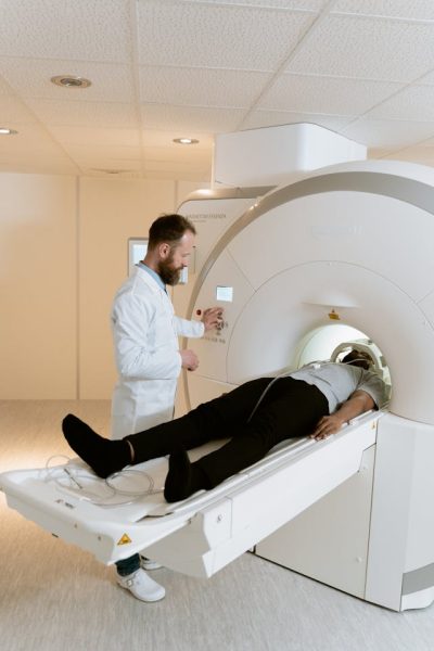
First, let’s explore mri in salt lake city. MRI, or magnetic resonance imaging, is the most advanced of the two. It was developed in the 1970s and became mainstream about a decade later. MRI machines use powerful magnets to create strong electromagnetic fields. Those fields are actually around 30,000 times stronger than Earth’s magnetic field. At this point, it’s important to mention that our bodies are mostly made of water. Water contains hydrogen atoms that have protons that act like tiny spinning magnets themselves.
Under normal circumstances, the hydrogen atoms in our bodies point in various directions and create random patterns. When we’re exposed to the strong electromagnetic fields created in MRI machines, though, the hydrogen atoms align with them, much like the needle in a compass does with Earth’s magnetic field. Then, MRI machines send out strong radio frequency pulses that disrupt the alignment of the hydrogen protons. When the pulses stop, the protons realign. This process causes them to release energy.
MRI machines detect the signals our hydrogen protons give off. Each type of tissue in our bodies contains different amounts of water and has a different composition. As such, they all give off different types of signals during MRI scans. MRI machines use mathematical algorithms to interpret those varying signals and generate clear, very detailed pictures of the inner workings of the body.
What Are MRIs Used for?
MRIs can distinguish different types of tissues, and they can clearly show all of them in the images they produce. That makes them useful for many purposes. They’re particularly helpful in soft tissue imaging. Doctors often use them for imaging organs, the brain and spinal cord, ligaments, tendons, and other soft tissues. They can certainly be used for bones as well, though. While the thought of a machine affecting the alignment of atomic particles in our bodies may seem disturbing, MRIs are completely safe.
Taking a Closer Look at X-Rays
Now, let’s take a closer look at X-rays. This technology has been around much longer than MRI. It dates back to the late 1800s. For quite some time, it was the only non-invasive way for doctors to look inside the human body. It paved the way for MRI, CT, PET, ultrasound, and other medical imaging technologies.
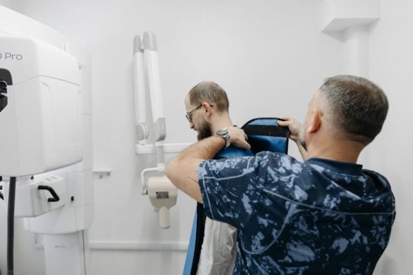
X-ray machines work by generating strong fields of high-energy electromagnetic radiation. That radiation can easily pass through most objects, including the human body. It doesn’t realign our hydrogen protons, though. Instead, it passes through different types of tissues at different rates. A film or digital detector picks up those rays when they pass through us to create images.
Since X-rays pass through tissues at varying rates, they cause them to look different in medical images. Dense materials, like bones, appear white. Less dense tissues, like organs absorb fewer X-rays, so they appear gray. Air-filled areas, such as the lungs, absorb very few X-rays, so they appear dark. Those differences allow doctors to distinguish between tissues. At the same time, X-ray images highlight abnormalities, which allows doctors to see them.
How Are X-Rays Used?
Images produced by X-rays aren’t quite as clear as those from MRIs. With that being the case, they can’t be used to detect as many medical problems and conditions. Still, they excel in certain areas. They’re effective for detecting broken bones and joint issues like arthritis. They can pinpoint cavities in our teeth and show infections, such as pneumonia. They can also alert doctors to certain types of tumors and other anomalies. In short, dark patches in areas that should be light or light patches in those that should be dark are often warning signs of issues that doctors should consider.
It’s worth mentioning that the radiation used in X-rays can potentially cause damage to our cells, which may increase the risk of developing cancer. Radiologists take every possible precaution when performing X-rays, though. They provide patients with lead aprons to prevent radiation exposure to parts of the body that aren’t being examined. Additionally, exposure to radiation is only occasional and very short-lived. With that being the case, the dangers are minimal, and the benefits of X-rays far outweigh the risks.
Benefiting From Medical Imaging
Medical imaging has brought about a long list of benefits. It’s a non-invasive way to see inside the human body, and it has enabled doctors to diagnose and treat a wide range of medical issues. It has saved countless lives over time while also saving patients a great deal of pain, suffering, and grief from ineffective treatments. It also allows doctors to catch and treat many conditions early on before they have a chance to grow worse.
MRI and X-ray are very different technologies. Each one serves its own purposes, and both offer several advantages. Though MRI provides more detailed images, X-ray still has its place in the medical field. In general, MRI is considered the safest of the two because it doesn’t involve radiation exposure. When used appropriately following proper safety protocols, though, the potential risks associated with X-rays are minimal.